One of the applications that has received the most attention in the latest years is the segmentation of organs or pathological tissues. Computational segmentation methods not only facilitate the demarcation of tissues from noisy and complex data, but it also allows rapid, automatic and repeatable extraction of geometric parameters (diameter, area, volume, shape, …) of great help for patient diagnosis and monitoring.
Among the multiple existing segmentation methods, deformable models have stand out due to their combined capability for regularization, by means of local shape constraints, and incorporation of prior knowledge. They are dynamic models that, starting from an initial state, are able to evolve until they fit the low level image features, with ability to manage topological changes, as observed in the following image. Additionally, they are suitable for tracking of deformable objects in video sequences.
Gradiant has personnel with experience in digital medical image processing and analysis, both from 2D and 3D data (CT, MRI, SPECT, …), and in particular, in 3D segmentation with deformable models. We have worked with geodesic models and model initialization using multiresolution features. Our background in the field of pattern recognition can be applied to the most recent segmentation techniques, which combine deformable models with region classification methods based on appearance statistical models.
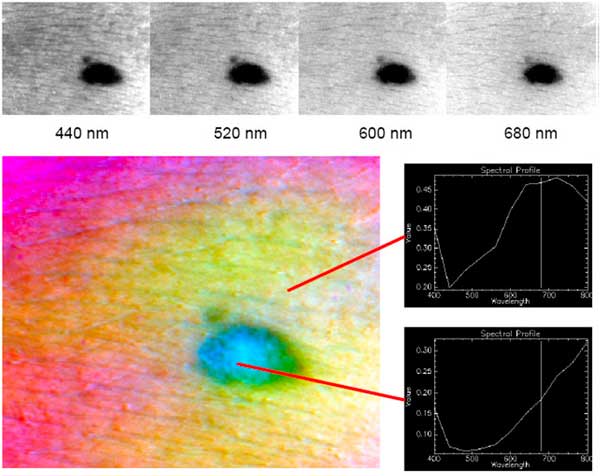
Besides, our experience in multi- and hyper-spectral image acquisition and analysis is also of application to medical image segmentation, especially in dermatology, where this image modality is being successfully used for detection and monitoring of melanoma. The analysis of the information provided by the different frequency bands allows revealing pathological areas that are not observable in the visible range, like the area tagged in yellow in the image.